Harness the full potential of your digital pathology data

A containerized web-based platform for the analysis, visualization, management and annotation of whole-slide digital pathology imaging data

What is DSA?

The Digital Slide Archive (DSA) is a platform that provides the ability to store, manage, visualize and annotate large imaging data sets. The DSA consists of an analysis toolkit (HistomicsTK), an interface to visualize slides and manage annotations (HistomicsUI), a database layer (using Mongo), and a web-server that provides a rich API and data management tools (using Girder). This system can:

  • Organize images from a variety of assetstores, such as local files systems and S3.
  • Provide user access controls.
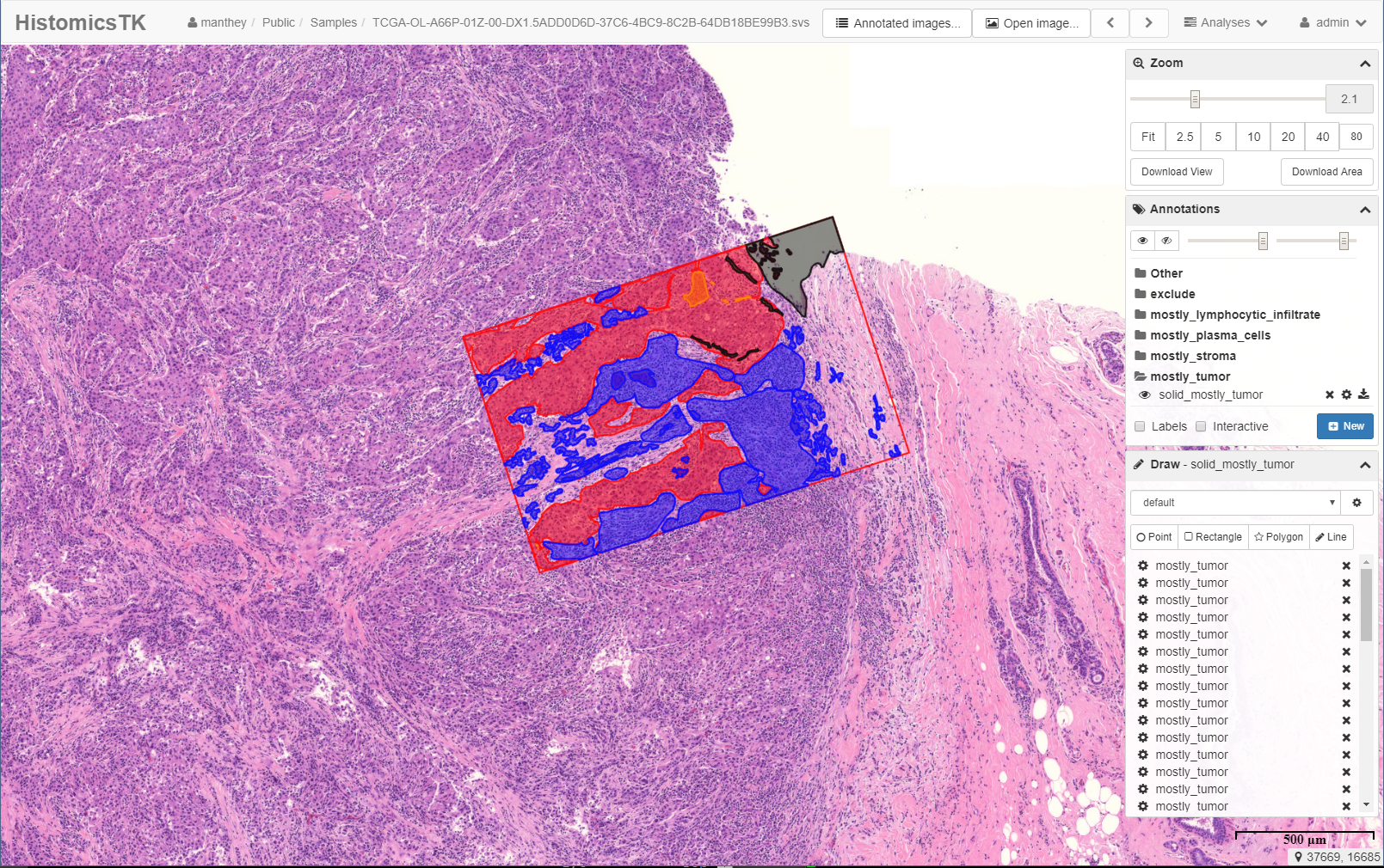
  • Image annotation and review.
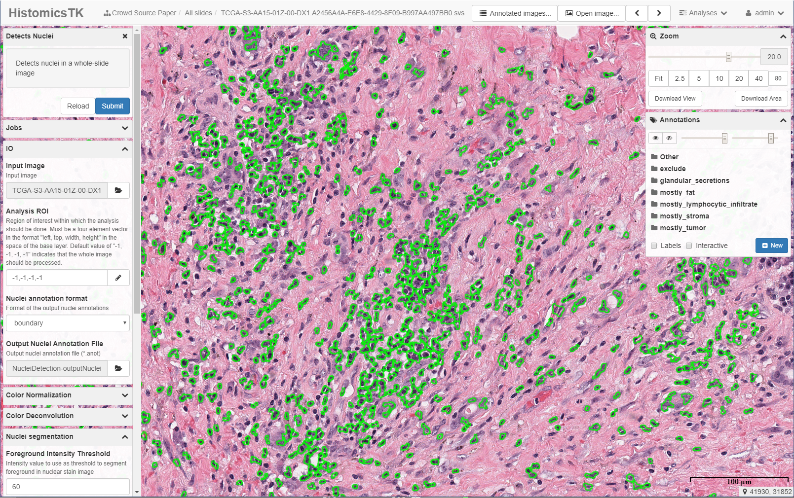
  • Run algorithms on all or parts of images.

HistomicsUI

HistomicsUI is a web-based application for examining, annotating, and processing histology images to extract both low and high level features (e.g. cellular structure, feature types).

Secure Data Management

The DSA provides fine-grained user or role-based access to datasets, images & metadata, and annotations. Amazon S3 hosting supported.

RESTful APIs

A rich API allows programmatic control over users, data, annotations, and algorithms, enabling automation of DSA tasks and integration with other tools and platforms.

Visualization and Annotation

An optimized user interface provides fluid exploration of large whole-slide images and tools for efficient generation of image markups.

Execution Engine

Girder provides distributed execution and monitoring of algorithm and analytics jobs.

Broad Support for Histology Image Formats

A wide variety of tiled image formats are supported, including tiff, svs, and jp2. Images can be retiled automatically as needed for processing algorithms. Additional formats can be added with a pluggable Python interface.

HistomicsTK

HistomicsTK is a Python image-processing toolkit for quantitative analysis of whole-slide digital pathology images.

Preprocessing

HistomicsTK provides color normalization and deconvolution operations to improve the robustness of analytic pipelines.

Object Detection and Segmentation

HistomicsTK contains a number of classical image analysis and machine-learning based algorithms for object detection and segmentation of subcellular structures and tissues.

Feature Extraction and Predictive Modeling

Object and patch-level features describing shape, texture, and color can be used to build machine-learning models.

Extensibility

Users can integrate their custom algorithms through a containerization process that auto-generates DSA user-interfaces.

Broad Support for Histology Image Formats

The same wide variety of histology images that can be viewed can be used with any processing algorithms. Sub-images can be processed at custom tile sizes and magnifications as needed.

Success Stories

View Stories

Papers & Publications

Read Papers
Lee A.D. Cooper, PhD
HistomicsTK Lead Associate Professor of Pathology
David A. Gutman, MD, PhD
Digital Slide Archive Lead Associate Professor of Neurology
David Manthey
Software Engineering & Deployment Staff R&D Engineer